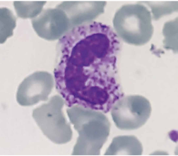
Granulaciones tóxicas: Un marcador clave en sepsis
DOI:
https://doi.org/10.37711/rpcs.2025.7.1.563Palabras clave:
sepsis, gránulos citoplasmáticos, biomarcadores, granulaciones tóxicasResumen
Actualmente, la sepsis es considerada una disfunción orgánica potencialmente mortal que representa un desafío alarmante en la práctica médica, de manera especial en el manejo de pacientes en estado crítico. En este sentido, las granulaciones tóxicas en neutróflos han surgido como un marcador ideal para facilitar su diagnóstico. Por ello, el objetivo del estudio fue analizar la importancia de las granulaciones tóxicas en el diagnóstico de la sepsis, su capacidad como marcador pronóstico y la incorporación en la práctica clínica diaria. Se realizó una revisión bibliográfica de estudios publicados entre 2019 y 2024 que evaluaron la relación de las granulaciones tóxicas con la sepsis. Las granulaciones tóxicas se relacionaron con una respuesta inflamatoria severa, tal como la sepsis, pero también se han observado en otras infecciones graves y algunos trastornos hematológicos, limitando su capacidad como único marcador. En conclusión, las granulaciones tóxicas constituyen una herramienta diagnóstica, pronóstica y de seguimiento esencial en la sepsis, a pesar de que se necesita más investigación para estandarizar su uso en la práctica clínica.
Descargas
Citas
Kim M, Choi J. An update on sepsis biomarkers. Infect Chemother. [Internet]. 2020 [Consultado el 24 de noviembre de 2024];52(1):1–18. https://doi.org/10.3947/ic.2020.52.1.1
Zhang W, Zhang Z, Pan S, Li J, Yang Y, Qi H, et al. The clinical value of hematological neutrophil and monocyte parameters in the diagnosis and identifcation of sepsis. Ann Transl Med. [Internet]. 2021 [Consultado el 24 de noviembre de 2024]; 9(22):1680. Disponible en: https://atm.amegroups.org/article/view/83998/html
Meyer N, Prescott H. Sepsis and Septic Shock. N Engl J Med. [Internet]. 2024 [Consultado el 10 de abril de 2025]; 391(22):2133–2146. Disponible en: https://www.nejm.org/doi/full/10.1056/NEJMra2403213
Giamarellos-Bourboulis E, Aschenbrenner A, Bauer M, Bock C, Calandra T, Gat-Viks I, et al. The pathophysiology of sepsis and precision-medicine-based immunotherapy. Nat Immunol. [Internet]. 2024 [Consultado el 10 de abril de 2025];25(1):19–28. doi: 10.1038/s41590-023-01660-5
Othman A, Sekheri M, Filep J. Roles of neutrophil granule proteins in orchestrating inflammation and immunity. FEBS J. [Internet]. 2022 [Consultado el 24 de noviembre de 2024];289(14):3932–3953. https://doi.org/10.1111/febs.15803
Kim Y, Park H, Rhee H, Hong J, Han K. Neutrophils with Toxic Granulation Show High Fluorescence with Bis(Zn2+- ipicolylamine) Complex. Ann Clin Lab Sci. [Internet]. 2009 [Consultado el 10 de abril de 2025];39(2):114–119. Disponible en: http://www.annclinlabsci.org/content/39/2/114.long
Sahota P, Spaet R, Bentley P, Wojcinski Z. The illustrated dictionary of toxicologic pathology and safety science [Internet]. Primera edición digital. Florida: Editorial CRC Press; 28 de mayo de 2019 [Consultado el 10 de abril de 2025]. Disponible en: https://www.routledge.com/The-Illustrated-Dictionary-of-Toxicologic-Pathology-and-Safety-Science/Sahota-Spaet-Bentley-Wojcinski/p/book/9781498754712
Chander S, Sharma R, Sharma A, Verma P. Toxic granules in neutrophils in sepsis patients: Does it really helpful? Muller J Med Sci Res. [Internet]. 2023 [Consultado el 24 de noviembre de 2024];14(1):19–22. Disponible en: https://journals.lww.com/mjmr/fulltext/2023/14010/toxic_granules_in_neutrophils_in_sepsis_patients_.5.aspx
Van de Vyver A, Delport E, Esterhuizen M, Pool R. The correlation between C-reactive protein and toxic granulation of neutrophils in the peripheral blood. S Afr Med J. [Internet]. 2010 [Consultado el 10 de abril de 2025];100(7):442–
Disponible en: https://www.ajol.info/index.php/samj/article/view/69671
Al-Gwaiz L, Babay H. The Diagnostic Value of Absolute Neutrophil Count, Band Count and Morphologic Changes of Neutrophils in Predicting Bacterial Infections. Med Princ Pract. [Internet]. 2007 [Consultado el 10 de abril de 2025];16(5):344–347. https://dx.doi.org/10.1159/000104806
Salgado D, Carvalho R, Oliveira M, Santos E, Brito L. The investigation of the presence of toxic granulation for septicemia hematologic diagnostic. Rev Bras Hematol Hemoter. [Internet]. 2007 [Consultado el 10 de abril de 2025];29(4):373–377. Disponible en: https://www.scielo.br/j/rbhh/a/H764dfZNnVkpN7NN5GkXztn/abstract/?lang=en&format=html
Kabutomori O, Kanakura Y, Watani Y. Induction of toxic granulation in neutrophils by granulocyte colony-stimulating factor. Eur J Haematol. [Internet]. 2002 [Consultado el 24 de noviembre de 2024];69(3):187–188. Disponible en: https://pubmed.ncbi.nlm.nih.gov/12406015/
Kabutomuri O. Toxic Granulation Neutrophils and C-Reactive Protein. JAMA Intern Med. [Internet]. 2000 [Consultado el 10 de abril de 2025];160(21):3326–3327. Disponible en: https://jamanetwork.com/journals/jamainternalmedicine/article-abstract/526460
Gigon L, Yousef S, Karaulov A, Simon H. Mechanisms of toxicity mediated by neutrophil and eosinophil granule proteins. Allergol Int. [Internet]. 2021 [Consultado el 24 de noviembre de 2024];70(1):30–38. Disponible en: https://pubmed.ncbi.nlm.nih.gov/33277190/
Faurschou M, Borregaard N. Neutrophil granules and secretory vesicles in inflammation. Microbes Infect. [Internet]. 2003 [Consultado el 24 de noviembre de 2024];5(14):1317–1327. Disponible en: https://pubmed.ncbi.nlm.nih.gov/14613775/
Huerto Aguilar JL, Martos Fustamante DL. Inclusiones azulverdosas en neutrófilos y monocitos: ¿resultados de riesgo crítico? Rev Peru Cienc Salud [Internet]. 25 de septiembre de 2023 [Consultado el 10 de abril de 2025];5(3):191-
https://doi.org/10.37711/rpcs.2023.5.3.424
Naeim F, Rao P, Song S, Phan R. Granulocytic Disorders. Atlas of Hematopathology: Morphology, Immunophenotype, Cytogenetics, and Molecular Approaches [Internet]. Segunda edición digital. Londres: Editorial Elsevier Science; 1 de febrero de 2018 [Consultado el 10 de abril de 2025]. Disponible en: https://www.callisto.ro/carte/atlas-of-hematopathology-morphology-immunophenotype-cytogenetics-and-molecular-approaches--i10094?lang=en
Feng P, He Y, Guan P, Duan C, Huang J, Chai Z, et al. Serum Procalcitonin, Hematology Parameters, and Cell Morphology in Multiple Clinical Conditions and Sepsis. J Clin Lab Anal. [Internet]. 2024 [Consultado el 24 de noviembre de 2024];38(19–20):e25100. https://doi.org/10.1002/jcla.25100
Bain B, Bates I, Laffan M. Hematología Práctica de Dacie y Lewis [Internet]. 12ª ed. Barcelona: Editorial Elsevier; 2018 [Consultado el 10 de abril de 2025]. Disponible en: https://www.sciencedirect.com/book/9780702066962/dacie-and-lewis-practical-haematology
Kroft S. Infectious diseases manifested in the peripheral blood. Clin Lab Med. [Internet]. 2002 [Consultado el 10 de abril de 2025];22(1):253–277. doi: 10.1016/s0272-2712(03)00074-x
Bahadur S, Kalonia T, Kamini K, Gupta B, Kalhan S, Jain M. Changes in peripheral blood in SARS CoV-2 patients and its clinico-pathological correlation: A prospective cross-sectional study. Int J Lab Hematol. [Internet]. 2021[Consultado el 29 de noviembre de 2024];43(6):1334-1340. Disponible en: https://pmc.ncbi.nlm.nih.gov/articles/PMC8653337/
Aro P, Paredes R, Sánchez J, Estrada S, Reyes S, Tokumura C. Alteraciones en los parámetros hematológicos y anormalidades morfológicas en células sanguíneas en pacientes hospitalizados con COVID-19. Rev Hematología [Internet]. 2023 [Consultado el 29 de noviembre de 2024];27(2):20–28 Disponible en: https://revistahematologia.com.ar/index.php/Revista/article/view/537/761
Refaat L, Abdellateif M, Bayoumi A, Khafagy M, Kandeel E, Nooh H. Detection of abnormal lymphocytes in the peripheral blood of COVID-19 cancer patients: diagnostic and prognostic possibility. Hematology [Internet]. 2022 [Consultado el 10 de abril de 2025];27(1):745–756. https://doi.org/10.1080/16078454.2022.2089830
Palacio M, Jaramillo P, Gálvez K. Hallazgos morfológicos en síndromes mielodisplásicos. Rev. Colomb. Hematol. Oncol. [Internet]. 2021 [Consultado el 6 de diciembre de 2024]; 8(1):62–75. https://doi.org/10.51643/22562915.117
Bain B, Ahmad S. Chronic neutrophilic leukaemia and plasma cell-related neutrophilic leukaemoid reactions. Br J Haematol. [Internet]. 2015 [Consultado el 10 de abril de 2025];171(3):400–410. doi: 10.1111/bjh.13600
Marín L, Martínez C. Tuberculosis congénita: a propósito de un caso. Rev Inst Med Trop. [Internet]. 2023 [Consultado el 29 de noviembre de 2024];18(2):51–61. https://doi.org/10.18004/imt/2023.18.2.8
Van der Poll T, Shankar-Hari M, Wiersinga W. The immunology of sepsis. Immunity [Internet]. 2021 [Consultado el 29 de noviembre de 2024];54(11):2450–2464. Disponible en: http://www.cell.com/article/S1074761321004490/fulltext
Laguado-Nieto M, Amaris-Vergara A, Vargas-Ordóñez J, Rangel-Vera J, García-León S, Centeno-Hurtado K. Actualización en sepsis y choque séptico en adultos. MedUNAB. [Internet]. 2019 [Consultado el 29 de noviembre de 2024];22(2):213– 227. https://doi.org/10.29375/01237047.3345
Alvarado J, Saquicela C, Nieto J, García D. Conceptos actuales de sepsis y shock séptico. J Am Health. [Internet]. 2020 [Consultado el 10 de abril de 2025];3(2):102–116. Disponible en: https://jah-journal.com/index.php/jah/article/view/38/78
Velly L, Freund Y. Biomarkers of sepsis: An old story or an exciting future? Emergencias [Internet]. 2022 [Consultado el 4 de diciembre de 2024];34(6):474–5. doi: 10.55633/s3me/E014.2022
Descargas
Publicado
Cómo citar
Número
Sección
Licencia
Derechos de autor 2025 Angel Isaias Pandashina Masabanda, María José Romero Pilacuan, Erika Alejandra Vega Ramos

Esta obra está bajo una licencia internacional Creative Commons Atribución 4.0.